Brain Mapping Progress
Brain mapping breakthrough

A new and relatively simple technique for mapping the wiring of the brain has shown a correlation between how well connected an individual’s brain regions are and their intelligence.
The following article first appeared in NR Times, the magazine which covers brain injury and brain conditions from every angle.
In recent years, there has been a concerted effort among scientists to map the connections in the brain – the so-called ‘connectome’ – and to understand how this relates to human behaviours, such as intelligence and mental health disorders.
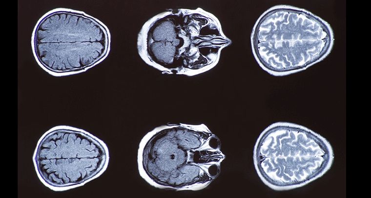
Now, in research published in the journal Neuron, an international team led by scientists at the University of Cambridge and the National Institute of Health (NIH), US, has shown that it is possible to build a map of the connectome by analysing conventional brain scans taken using a magnetic resonance imaging (MRI) scanner.
The team compared the brains of 296 typically-developing adolescent volunteers. Their results were then validated in a cohort of a further 124 volunteers.
The team used a conventional 3T MRI scanner (with ‘3’ representing the strength of the magnetic field); however, Cambridge has recently installed a much more powerful Siemens 7T Terra MRI scanner, which should allow this technique to give an even more precise mapping of the human brain.
A typical MRI scan will provide a single image of the brain, from which it is possible to calculate multiple structural features. This means every region of the brain can be described using as many as ten different characteristics.
Researchers showed that if two regions have similar profiles, they are described as having “morphometric similarity” and it can be assumed that they are a connected network.
They verified this assumption using publicly-available MRI data on a cohort of 31 juvenile rhesus macaque monkeys to compare to “gold-standard” connectivity estimates in that species.
Using these morphometric similarity networks (MSNs), the researchers were able to build up a map showing how well connected the ‘hubs’ – the major connection points between different regions of the brain network – were.
They found a link between the connectivity in the MSNs in brain regions linked to higher order functions – such as problem solving and language – and intelligence.
“We saw a clear link between the ‘hubbiness’ of higher-order brain regions – in other words, how densely connected they were to the rest of the network – and an individual’s IQ,” says Jakob Seidlitz, a PhD candidate at the University of Cambridge and NIH.
“This makes sense if you think of the hubs as enabling the ow of information around the brain – the stronger the connections, the better the brain is at processing information.”
While IQ varied across the participants, the MSNs accounted for around 40 per cent of this variation. It is possible that higher-resolution multi-modal data provided by a 7T scanner may be able to account for an even greater proportion of the individual variation, say the researchers.
“What this doesn’t tell us, though, is where exactly this variation comes from,” adds Seidlitz.
“What makes some brains more connected than others? Is it down to their genetics or educational upbringing, for example? And how do these connections strengthen or weaken across development?”
For more articles on brain injury and brain conditions see www.nrtimes.co.uk.
Copyright © 2019 UDAV - All Rights Reserved.